
ANATOMY OF THE HAND – simplified!
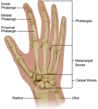 The skeleton of the hand is made up of 29 bones; there are 9 dice-sized carpal bones near the wrist, joined closely to each other, 5 tubular metacarpal bones that end at the knuckles and the phalanges of the fingers and thumb (3 in each finger and 2 in the thumb). Bones are joined together by ligaments; ligaments are strong fibrous bands that stabilise joints and stop the joint moving in directions that it is not designed to move. Ligaments can be torn completely or partly (a sprain).
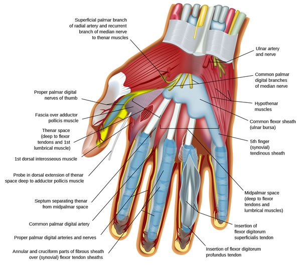
There are numerous small muscles in the hand itself that move the fingers and thumb. These small muscles form the soft pad at the base of the thumb and the equivalent pad on the little finger side of the hand. The most important and strongest muscles that straighten and bend the fingers and thumb are situated in the forearm. Muscles are joined to bone by tendons which are the sinewy cords that you may be able to see on the back of your hand and the front of your wrist. The muscles that bend the fingers are called flexors and those that straighten the fingers are called extensors. The tendons attach to the phalanges in the fingers and thumb. The large number of muscles in the hand allow the very delicate and precise movement that is possible. Skin covers the hand; the skin on the palm of the hand is thick and not particularly stretchy, the skin on the back of the hand (dorsum) is thin and pliable. There are two arteries that carry blood to the hand, the radial and ulnar arteries. The radial artery can be felt as the pulse at the wrist. There are three nerves that pass to the hand, the median, ulnar and radial nerves. Nerves have two functions, they tell you what you are feeling (sensation) and carry messages from the brain to muscles to cause a movement.